I promised not to post this report before the item shown below was published in The New Energy Times by Steven Krivit.
http://newenergytimes.com/news/2007/NET21.htm
I hope that experiments now in progress will bring more clarity into the SPAWAR effect discovered by Stanislaw Szpak et al. They have been studying several codeposition
effects for more than ten years. But their use of CR-39 detectors is relatively recent.
Click to see the list of links
319) A contribution to Galileo Project
Ludwik Kowalski; 2/25/2006
Department of Mathematical Sciences
Montclair State University, Upper Montclair, NJ, 07043
1) Introduction
As described in the unit #314, I volunteered to be part of the The Galileo Project (TGP) organized by Steven Krivit. Before reading beyond this introduction,
please go to
http://newenergytimes.com/news/2006/NET19.htm
and click the link to item #7, entitled "Extraordinary Evidence." You also should read item #8; I do not want to repeat what has already been described on the above
webpage. Read these two items carefully and then read what is below. This unit is about our attempt to replicate one of the recent SPAWAR experiments. The study was
conducted together with Michael Stolarz, a physics major at Montclair State University, and with two colleagues -- Brian Scanlan and Simon Young. Michael will present a
separate paper at the Sigma Xi student research conference. Richard Oriani, who lives in Minneapolis, was our adviser. It is important to emphasize that several
papers about nuclear processes triggered by chemical processes in electrolytic cells have already been published by the SPAWAR team in professional journals (see
references in 1). The experiment we attempted to replicate has also been published (6).
The heavy-water electrolyte we used was identical to that described in (1). The time table for the electrolysis (see below) was also very similar. We were able to
replicate SPAWAR results that were presented, by Frank Gordon (2). But our interpretation of results is different from that offered by SPAWAR team. The diameters of
dominant dark pits, on our CR-39 detector, were found to be too large to be attributed to to alpha particles, or to smaller nuclear projectiles.
Inserted on 3/3/07--> After finishing writing this unit, three days ago, I decided to make a presentation at the upcoming APS meeting. I wrote to the session
chairman, Scott Chubb, that I have something important to share and would be happy to take place of someone who does not show up. He offered me a time slot and suggested
that I write an abstract and distribute it at the conference. Here is my abstract”
Title: Nuclear or not nuclear? That is the question.
Authors: Ludwik Kowalski, Brian Scanlan and Simon Young.
Text: An experiment described by Frank Gordon, last summer (1), has been successfully replicated at Montclair State University. Copious pits were observed on the
CR-39 chip that was in contact with the silver cathode wire in the magnetic field. No such pits were observed on the identical chip from the cell that was not in magnetic
field. The two cells were in series with each other during the electrolysis. Diameters of large post-electrolysis pits have been compared with diameters of pits due to
alpha particles from a radioactive source. On the basis of this analysis, and on the basis calibration curves reported by Roussetski et al. (2), we conclude that large
pits on our CR-39 chip, from one experiment, cannot possibly be attributed to alpha particles, or to less massive nuclear projectiles.
References:
1) F. Gordon et al, at the Naval Science & Technology Partnership conference hosted by the National Defense Industrial Association and the Office of Naval Research
(Washington, DC, August 2, 2006). ) See items 7 and 8 in
2) A.S. Roussetski, A.G. Lipson and V.P. Andreanov; "Nuclear emission from titanium hydride/deuteride induced by powerful picisecond laser beam." Proceedings of the
10th international conference on cold fusion, pages 559-566.
2) Experimental Method
Two identical electrolytic cells were connected to power supply, as shown in Figure 1. The little boxes, at the upper left corner represent integrated circuits designed
to maintain constant currents. To change the current we simply had to replace one box by another. The voltage of the power supply was sufficiently high (up to 50 V) to
support this mode of operation. This inexpensive method of keeping the current constant was introduced to us by Dr. Winthrop Williams. The experiment would not be possible
without his help in dealing with numerous practical problems.
- - - - - - - - - - - - - - - - - - - - - - - - - - - - - - - - - - - - - -
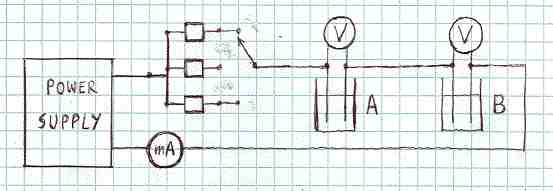
FIGURE 1
Our electric circuit contained nine integrated circuits (LM334 for lower currents and LM317T for higher currents). Only three of them are show in this diagram. A third
cell was connected in series with A and B. It is not shown in this figure because it was part of another experiment (to be described in another unit).
- - - - - - - - - - - - - - - - - - - - - - - - - - - - - - - - - - - - - -
The two electrolytic cells, like those described in (1), were made from acrylic. Each has a wire-made platinum anode and a wire-made silver cathode, as shown in Figure 2.
A CR-39 detector of nuclear particles was mechanically applied to the silver wire of each cell.
The only difference between the two cells was that one (A) was placed in a magnetic field crated by two very strong neodymium magnets while the other (B) was far away
from any source of magnetic field higher than terrestrial.
- - - - - - - - - - - - - - - - - - - - - - - - - - - - - - - - - - - - - -

FIGURE 2
The distance between the anode and the cathode was approximately 15 mm. The height of the acrylic cell was 80 mm. The direction of the magnetic field lines (in cell A only)
was from the anode toward the cathode. The magnetic field near the cathode was close to 0.2 T.
- - - - - - - - - - - - - - - - - - - - - - - - - - - - - - - - - - - - - -
The following time table was used during the electrolysis:
Current 0.11 mA, 24 hours
Current 0.21 mA, 24 hours
Current 0.56 mA, 12 days, till the electrolyte changed from brown to transparent
Current 1.0 mA, 24 hours
Current 5.0 mA, 24 hours
Current 10.1 mA, 24 hours
Current 26.9 mA, 24 hours
Current 55 mA, 24 hours
Current 127 mA, 24 hours
Heavy water had to be added to cells in order to compensate for what was lost due to evaporation and electrolysis. Increases in electric current were accompanied by
increases of potentials across the cells. The potentials at the beginning of the electrolysis were 0.96 volts on each cell; the potentials at the end of the
electrolysis were close to 8 volts. (At 1 mA they were close to 1.6 V and at 50 mA they were close to 5.5 V).
After the electrolysis the CR-39 chips were removed and visually examine. The opalescencent lines were seen on the chip from the cell A, more or less where
the cathode wires were. No such lines were seen on the chip from the cell B. Another detail is worth mentioning. Before the electrolysis the cathodes in both cells
were in contact with detectors along the entire length. After the electrolysis the cathode wire in cell B was still in contact with the detector along the entire
length. But the cathode wire in the cell A bulged away from the detector, except near the edges. The chips ere etched together for
3 hours in the standard NaOH electrolyte (6.25 N and 68 C), rinsed and examined under the microscope. The CR-39 chip removed form the cell A had several areas in
which numerous pits were observed, as illustrated in Figure 3. In that respect our results are in very good agreement with results reported by the SPAWAR team. The
CR-39 chip removed from the cell B had no areas covered with such pits. That observation is also consistent with what was reported by the SPAWAR team. We agree with
them that the magnetic field plays a role in the formation of pits. The mechanism of this effect is not clear at this time.
- - - - - - - - - - - - - - - - - - - - - - - - - - - - - - - - - - - - - -
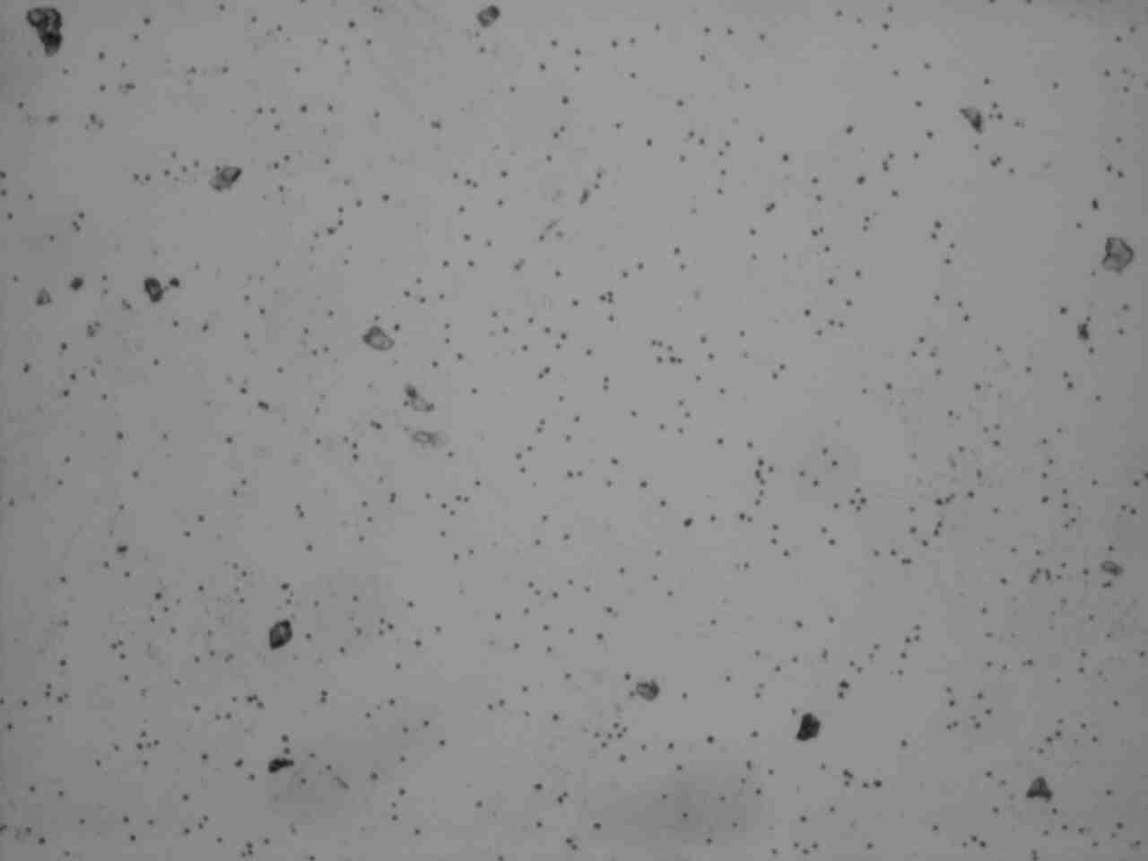
FIGURE 3A
Dot-like pits due to alpha particles from our Am-241 source at magnificatin 40. Surface defects or dust can also be seen. The area on this low magnification photo
is 1.3 by 0.9 mm.
- - - - - - - - - - - - - - - - - - - - - - - - - - - - - - - - - - - - - -
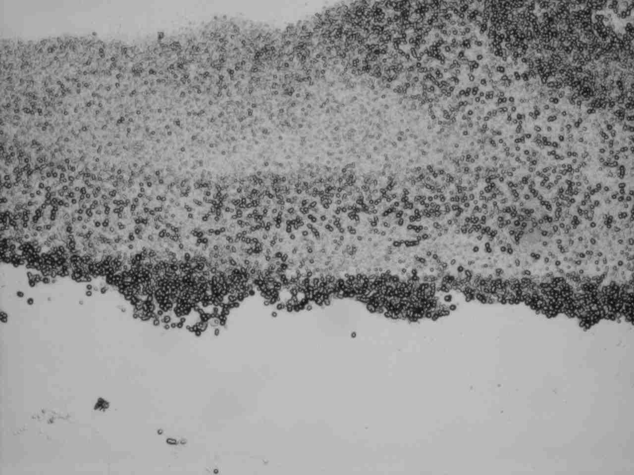
FIGURE 3B
Piits on the post-electrolysis chip from the cell A. Etching conditions and magnification were the same as for the photo on Figure 3A. The area on this low magnification
photo is 1.3 by 0.9 mm. Next four figures should be used to compare diameters of post-electrolysis pits with diameters of pits due to alpha particles.
The claim of nuclear origin of pits, made by SPAWAR team, was based on comparisons of observed pits with pits that were known to be due to alpha particles. We followed the
same approach. A fresh CR-39 chip was irradiated with alpha particles from an 241 Am source. It was etched together with the post-electrolysis chips. Figure 4 shows pits
due to alpha particles, under the microscopic magnification of 200. Figure 5 shows pits on the chip removed from the cell A, under the same magnification. It is clear that
the post-electrolysis pits are larger than pits due to alpha particles. The same conclusion can be reached by comparing pits in Figures 6 and 7. They were taken under the
magnification of 400.
- - - - - - - - - - - - - - - - - - - - - - - - - - - - - - - - - - - - - -

FIGURE 4
Pits which are known to be tracks of alpha particles, under magnification of 200
- - - - - - - - - - - - - - - - - - - - - - - - - - - - - - - - - - - - - -
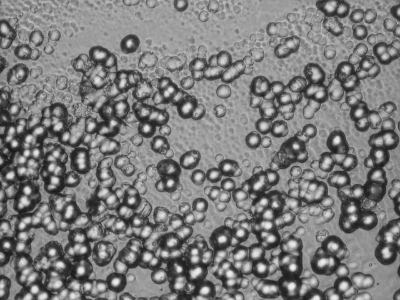
FIGURE 5
Pits on the post-electrolysis chip from the cell A. Etching conditions and magnification were the same as for the photo on Figure 4. Note that diameters of faint pits are
about the same, and smaller, than diameters of dark pits in Figure 4. Large dark pits can’t be due to alpha particles, or lighter projectiles.
- - - - - - - - - - - - - - - - - - - - - - - - - - - - - - - - - - - - - -
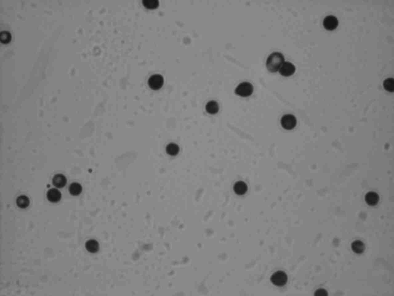
FIGURE 6
Pits which are known to be tracks of alpha particles, under magnification of 400
- - - - - - - - - - - - - - - - - - - - - - - - - - - - - - - - - - - - - -
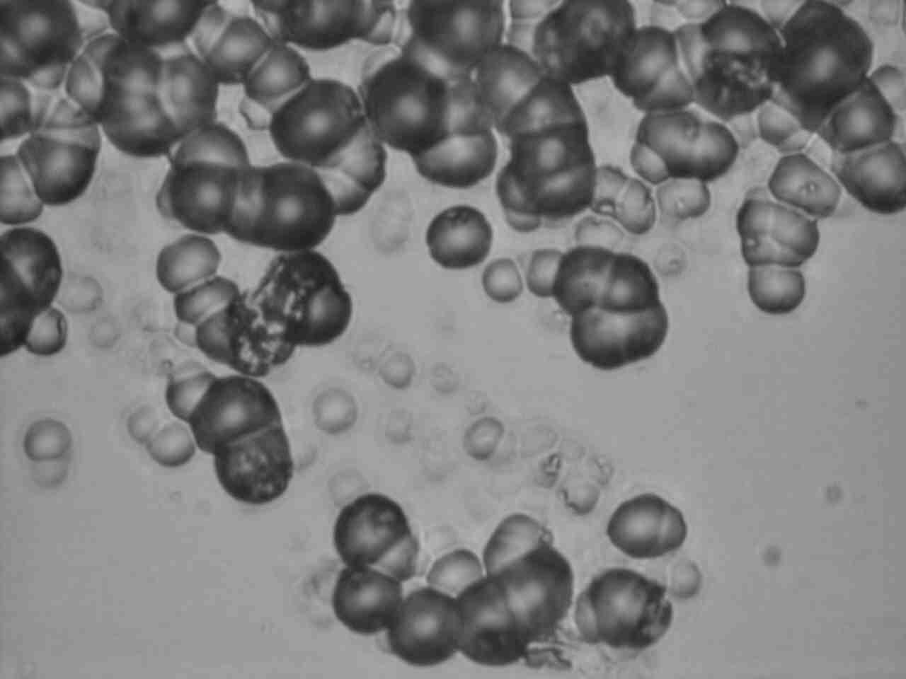
FIGURE 7
Pits on the post-electrolysis chip from the cell A. Etching conditions and magnification were the same as for the photo on Figure 6. Note that diameters of faint pits are
about the same, and smaller, than diameters of dark pits in Figure 6. Large dark pits can’t be due to alpha particles, or lighter projectiles.
- - - - - - - - - - - - - - - - - - - - - - - - - - - - - - - - - - - - - -
Absence of pits on the post-electrolysis chip B was a strong indication that the pit-formation process was strongly influenced by the magnetic field. That fact reminded us Marie Curie’s attempts to influence radioactivity by magnetic fields. These attempts, like those undertaken by other investigators, were not successful. Magnetic fields do influence some atomic processes but their ability to either slow down or to speed up emission of alpha particles (or protons) from atomic nuclei has never been demonstrated.
3) Discussion
Differences between 241Am pits and pits found on the post-electrolysis chips are not limited to sizes (typical alpha particles tracks are about 2.5 times smaller than typical post-electrolysis pits). Another difference becomes obvious when one tries to refocus the microscope under a larger magnification, such as 200 or 400. Isolated post-electrolysis pits seem to be more shallow than pits produced by alpha particles. Most post-electrolysis chips appear so close to each other that the individual boundaries are no longer identifiable. The areas covered by overlapping pits are whitish, as if they were affected by some kind of chemical corrosion.
Let us assume that individually identifiable post-electrolysis pits are due to nuclear particles, as claimed by the SPAWAR team (6), and not to a chemical, thermal or mechancal effect. What kind of particles can produce pits shown in Figure 3? To answer this question we will use results of measurements made by Roussetski et al. (4). These Russian scientists exposed CR-39 chips to protons and alpha particles of various energies. Then they etched the chips, measured the diameters and summarized the results in the form of a very useful graph. The smallest diameter (5 microns) was for protons of 3 MeV, the largest diameter (12 microns) was for alpha particles of 2 MeV. The etching in the NaOH (6N, 70 C) took 7 hours.
Our etching conditions were quite different, as specified in section 2. For that reason we decided to modify the original graph slightly, as shown in Figure 8. Our diameters are expressed in arbitrary units named prots. One prot, by definition, is the diameter due to 3 MeV protons, no matter how many microns it is under different etching conditions. The advantage of that representation is obvious; it decouples essential information (relative sizes of pits) from the laboratory-specific details about etching. The modified graph is likely to be valid, at least approximately, when etching conditions are not exactly the same as those used in (4).
- - - - - - - - - - - - - - - - - - - - - - - - - - - - - - - - - - - - - -
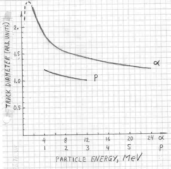
Relative diameters of tracks due to alpha particles and protons of various energies. The dashed line, at the upper left corner, was added to indicate that alpha particles cannot possibly produce tracks larger than about 2.5 prots. This expectation is based on the well known fact that alpha particles at energies below 2 MeV tend to capture electrons when they slow down. The dE/dx values do not increase when E becomes sammler than about 2 MeV.
- - - - - - - - - - - - - - - - - - - - - - - - - - - - - - - - - - - - - -
According to Figure 8, the mean diameter of alpha particles from 241Am (~ 5 MeV) is 1.7 prot. According to Figures 6 and 7, individually identifiable
post-electrolysis diameters are about 2.5 times larger than those due to 241Am. In other words, their average pit diameters are 1.7*2.5=4.5 prots This at
once rules out a possibility that the post-electrolysis pits are due to protons or to neutrons; diameters of tracks due to these particles would be smaller than 1.5 ports.
It also rules out a possibility that poste-electrolysis chips are due to alpha particles; diameters of these would be smaller than 2.5 prot. The only available avenue,
for defending nuclear origin of post-electrolysis chips, it to assume that they are caused by nuclear projectiles that are much heavier than alpha particles. Fission
fragments from a californium source, for example, were shown (5) to produce pits that were about 2.5 times larger than those due to alpha particles. But that does not
mean that any large pit on the CR-39 surface is a track of a massive nuclear projectile. An additional evidence would be needed to defend such claim.
According to (6), "the density of tracks registered by a CR-39 detector was found to be of a magnitude that provides indisputable evidence of their nuclear origin."
Our analysis of diameters of pits does not support this interpretation, unless only massive particles are responsible for the large pits shown in Figures 5 and 7.
A more likely explanation is that post-electolysis pits are due to a non-nuclear process taking place near the cathode. This kind of interpretation is supported by
presence of glossy regions on the chip from the cell A, visible before the chip was etched (see section 2). Prolonged exposures of CR-39 to alpha particles, undertaken
to produce overlapping pits, did produce foggy (but not glossy) areas on chips, but such areas could be seen only after etching. One might speculate, for example,
that hydroxyl ions, produced by reduction of water, somehow concentrated near the CR-39 surface, attacked the plastic.
The most puzzling fact, discovered by the SPAWAR team (6), and confirmed by our experiment, is that the unexplained pits are produced only when magnetic field is present.
Suppose the post-electrolysis pits are due to a non-nuclear process of some kind. In that case the role played by magnetic field is also far from being clear. As far as
we know, magnetic fields do not drastically change rates of chemical reactions. If this is true then something new, and totally unexpected, has been discovered by the
SPAWAR team. The magnetic field effect is interesting and worth investigating, even if pits are not of nuclear origin. The magnetic field effect, by the way, is a very
good indication that observed pits are not due to a familiar process, such as radioactive contamination in the cell or an ordinary chemical attack.
The effect of the electric field, reported in (2), is even more
puzzling. The difference of potential of 6000 volts, applied to parallel copper plates situated outside the cell with acrylic walls, should make a negligible contribution
to the electric field inside a cell with the electrolyte. This is due to the very high resistivity of acrylic in comparison with the low resistivity of the electrolyte. And
yet, the removal of an extremely small electric field stops a process by which track-looking pits are formed on the CR-39 detector. This effect is also interesting and
worth investigating.
Additional reflections. (Inserted on 3/16/07):
Why do many scientists reject the idea that a nuclear process can be triggered by a chemical process? Because this idea conflicts with the atomic model they have already
accepted. That model, progressively evolved during the 20th century, makes sense out of a large number of experimental facts. It states that chemical reactions are changes
at the level of the outermost electrons orbiting around atomic nuclei. Nuclear reactions, on the other hand, are changes occurring in tiny central regions of atoms.
Nuclear force bounding protons and neutrons in these tiny regions have very short ranges; they have no effect on orbiting electrons. Coulomb forces between electrons
and protons are negligibly small in comparison with nuclear forces. That is why electronic rearrangements (chemical changes) are generally expected to have negligible
effects on what is going on inside atomic nuclei.
In research, and in teaching, we progress from what is already known toward what remains hidden. New models of reality are refinements of models that are known to be
consistent with experimental facts. That is why many scientists say that cold fusion claims are extraordinary. They feel that one has to be extraordinary reserved toward
such claims. To “be reserved,” however, does not mean to “reject.” It means to ask many more questions than in the case of claims that are consistent with accepted models.
It is important to emphasize that that SPAWAR scientists, like many other cold fusion researchers, are experimentalists. They observe what happens and try to interpret it
in terms of familiar concepts. Our photos (shown in Figures 3, 5 and 7) are very similar to their photos (shown in reference 2). But we do not know if large pits on their
post-electrolysis chips are also considerably larger than pits on their chips exposed to alpha particles (under identical etching conditions). The disagreement is about
the idea that large post-electrolysis chips can be attributed to alpha particles, and lighter nuclear projectiles. Fortunately, experiments seem to be reproducible. In
that situation the disagreement can easily be turned into an agreement, one way or another. It becomes a simple matter of remeasuring, or repeating the experiment.
Will the clear picture emerge before the next week anniversary of cold fusion? I hope so.
Inserted 3/23/07
Our paper was discussed extensively on the private list for CMNS researchers. Replying to one message I wrote:
From past experience (placing CR-39 on a Pu-Be neutron source) I know that most tracks due to neutrons (via collisions with hydrogen nuclei in that material
C12-H18-O7) have diameters that are clearly smaller than diameters due to
alpha particles from the Am-241 source. Less than one percent of tracks due to neutrons had diameters comparable to those seen on the chip exposed to Am-241. I
attributed these large pits to the (n,alpha) reactions. That is why I said that pits larger than those due to Am-241 (identical magnification and etching) cannot be
attributed "to neutrons." Perhaps I should have said "to protons produced by neutrons." I am not denying a possibility that large dark pits on
our photos might be due "to carbon or oxygen ions produced by neutrons," as suggested at the APS meeting by Larry.
Let me estimate the number of neutrons that would be responsible for large pits Larry observed on the “back side” of his CR-39 chip. The rule of thumb
used by Frank Gordon (private communication during the APS conference) is that only one out of hundred thousand fast neutrons, passing through a CR-39 detector,
interacts with with atomic nuclei insid the detector. Taking this for granted, let me estimate the number of neutrons from the number of large pits. To accomplish this
I will make the following assumptions (which can easily be modifies). To accomplish this I will make four simplifying assumptions. These arbitrary assumptions can be
modified, if necessary.
a) One half out of 100,000 intercepted neutrons is colliding with heavy nuclei in CR-39 producing ions of C and O. The other half is scattered on hydrogen.
b) The mean range of heavy ions in CR-39 is 1 mg/cm2, which is 8 microns.
c) Larry’s large dark pits are due to only 25% of heavy ions produced in the last 8 microns of CR-39.
d) The thickness of the CR-39 chip is 1 mm, which is 1000 microns.
Thus the fraction of ions responsible for Larry’s pits is 0.25*8/1000=1/500. In other words, the number of large pits will be 500 times smaller than the number
of neutrons interacting with heavy nuclei inside the CR-39 chip. If 100,000 neutrons produce one interaction then 500*100000=5*107
neutrons will produce one Larry’s pit. For 1000 pits the number of neutrons would be 5*1010. This is 50 billion. Another
conclusion is that large pits should also be observed on the front surface of the CR-39 chip.
This speculation was posted on the private list for CMNS researchers. Here is a comment from one researcher: “ If 50 million neutrons were emitted by the cell
during the last 48 hours of the run when the current is at its highest value (and presumably making the nuclear reactions happen), the flux onto the CR-39 chip would
be 290,000 neutrons/sec. That'd make any neutron detector sing, so to speak. Also, assuming the neutrons are 2.5 Mev, it only takes 20 neutrons/cm^2/sec to make a
dose rate of 2.5 mrem/hr (1)....so the CR-39 chip would see a dose rate of ~36 rem/hour (assuming the chip was 1 cm2). The dose falls
off rapidly with distance but, still, such an experiment would be of considerable hazard to the health of bystanders. (1). Radiation Detection and Measurement, Glenn
Knoll, 1979, p. 802.”
After reading the 2002 paper of J.A. Frenje et al. (Review of Scientific Instruments, vol. 77, July 2002, p 2595) I realized that the assumption (a) above was
highly unrealistic. Experimental data show that the number of large pits due to heavy ions represents only a very small fraction of one percent of all pits, even for
14 MeV neutrons. For that reason the 50 million neutrons was underestimated by orders of magnitude. To produce 1000 pits on CR-39, the number of neutrons had to be
much larger than 5 billion. Fortunately, a Landauer neutron detector was used in one of The Galileo Project experiments. How many neutrons were actually detected
during that experiment? I will answer this question as soon as the outcome is announced.
References:
(1) Stanislaw Szpak · Pamela A Mosier Boss, Charles Young and Frank E. Gordon; “Evidence of nuclear
reactions in the Pd lattice.” Naturwissenschaften (2005) 00: 1–4 DOI 10.1007/s00114-005-0008-7. he document can be downloaded from the library at
http://lenr-canr.org
2) F. Gordon et al, at the Naval Science & Technology Partnership conference hosted by the National Defense Industrial Association and the Office of Naval Research
(Washington, DC, August 2, 2006). The document can be downloaded from the library at http://lenr-canr.org
3) D. Nikezic and K.N. Yu, “Formation and growth of tracks in nuclear track materials;” Material Science and Engineering R 46 (2004) p 51-123.
4) A.S. Roussetski, A.G. Lipson and V.P. Andreanov; “Nuclear emission from titanium hydride/deuteride induced by powerful picisecond laser beam.”Proceedings of
the 10th international conference on cold fusion, pages 559-566. The document can be downloaded from the library at http://lenr-canr.org
(5) D. Paul et al., “An SSNT study of spontaneous fission fragments from the soil gas samples of Bekerswar thermal springs;” Radiation Measurements 33 (2001)
p 167-169
6) Stanislaw Szpak, Pamela A Mosier Boss and Frank E. Gordon; “Further evidence of nuclear reactions in the Pd/D lattice.” Naturwissenschaften (2007)
DOI 10.1007/g00114-007-0221-7. Can be downloaded from the library at http://lenr-canr.org
Click to see the list of links